swiss medicaXXI century S.A.
Night blindness, a narrowing field of vision, and difficulty reading can slowly begin to affect everyday activities, even for people who remain under regular ophthalmic care. When these limitations persist, some begin exploring additional regenerative approaches.
Stem cell therapy for retinitis pigmentosa and diabetic retinopathy is one of several advanced biological approaches being investigated. However, the two diseases affect the retina in very different ways, which shapes the approach to care, the available evidence, treatment goals, and what can reasonably be discussed with each patient.
Understanding Retinitis Pigmentosa and Diabetic Retinopathy
Retinitis pigmentosa and diabetic retinopathy both affect the retina, the light-sensitive tissue at the back of the eye, but through distinct disease processes. As the underlying mechanisms, affected retinal structures, and disease progression are different, any discussion of additional treatment options must start with an accurate diagnosis and a thorough ophthalmic assessment.
Retinitis Pigmentosa Primarily Affects the Retina’s Light-Sensing Cells
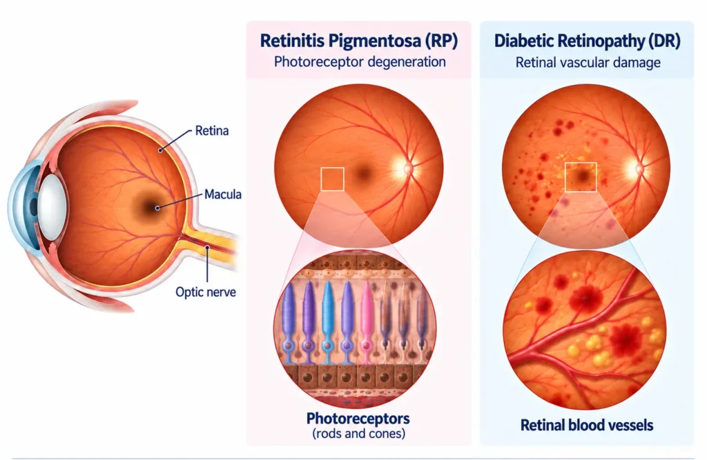
Retinitis pigmentosa (RP) is a group of inherited retinal disorders marked by the gradual break down of photoreceptors, the cells that respond to light. The retinal pigment epithelium (RPE), which supports and nourishes photoreceptors, may also be affected. For many patients, reduced vision in dim light is the first noticeable symptom. Peripheral vision may become increasingly restricted as the condition advances.
Because RP can result from many different genetic changes, the pattern and rate of vision loss can vary considerably between patients.
Diabetic Retinopathy Is a Disease of the Retinal Blood Vessels
Diabetic retinopathy develops when diabetes damages the small blood vessels that nourish the retina. These vessels can leak, close off, or lead to abnormal new vessel growth. Fluid may also accumulate in the macula, the central area of the retina responsible for detailed vision.
Why an Accurate Diagnosis Comes Before Any Treatment Decision
Blurred vision, floaters, and progressive visual field loss can show up across a range of very different retinal conditions, including retinitis pigmentosa, diabetic retinopathy, age-related macular degeneration (AMD), inherited retinal diseases, and inflammatory retinitis.
A proper diagnosis helps doctors find out what is causing the vision problem, which part of the retina is affected, and how much visual function remains. They may use several tests depending on the condition:
- Optical coherence tomography (OCT) is a scan that creates detailed images of the retinal layers. It can show thinning, swelling, fluid buildup, and other structural changes.
- Fundus photography takes pictures of the back of the eye. It helps doctors document visible changes in the retina and compare them over time.
- Visual field testing measures how much of the surrounding area a person can see while looking straight ahead. It is useful for detecting peripheral vision loss and tracking changes in the field of vision.
- Electroretinography (ERG) records how the retina responds to flashes of light. It helps assess how well the light-sensitive cells of the retina are functioning.
- Fluorescein angiography uses a fluorescent dye and a series of eye photographs to show blood flow through retinal vessels. It can reveal areas of leakage, blockage, or abnormal blood vessel growth.
- Genetic testing searches for changes in genes linked to inherited retinal diseases. It may help confirm the cause of conditions such as some forms of retinitis pigmentosa and determine whether gene-specific treatment or clinical trials may be relevant.
These findings help doctors distinguish between retinal conditions and choose treatment based on the specific disease process involved.
What Is the Standard Treatment for Retinitis Pigmentosa and Diabetic Retinopathy?
Standard treatment options differ substantially between retinitis pigmentosa and diabetic retinopathy. The following approaches do not apply equally to both conditions.
| Condition | Main treatment goal | Established care |
| Diabetic retinopathy | Reduce swelling and bleeding and protect vision | Diabetes management, anti-vascular endothelial growth factor (anti-VEGF) injections when needed, laser treatment, and surgery in selected cases |
| Retinitis pigmentosa | Preserve remaining vision | Regular retinal follow-up, low-vision rehabilitation, treatment of complications, and gene therapy in selected inherited cases |
Anti-VEGF Therapy, Laser, Surgery, and Genetic Care Have Different Roles
- Anti-VEGF injections help reduce retinal swelling and leakage from abnormal blood vessels. They are commonly used for diabetic retinopathy, particularly diabetic macular edema and proliferative disease, but are not a standard treatment for RP.
- Laser treatment may be used in diabetic retinopathy to treat leaking blood vessels or reduce the risk of complications from abnormal vessel growth. It is not used to treat the underlying retinal degeneration in RP.
- Eye surgery, most often vitrectomy, may be needed in advanced diabetic retinopathy to manage bleeding within the eye or tractional retinal detachment caused by scar tissue. It does not treat the underlying cause of RP, although surgery may occasionally be needed for separate eye complications.
- Gene therapy may be an option for selected inherited retinal diseases, including certain genetic forms of RP, when a treatment is available for the specific disease-causing variant. It is not a treatment for diabetic retinopathy.
Retinal Specialist Follow-Up Remains Essential
Retinal disease can progress without causing obvious new symptoms. Regular follow-up is therefore important for identifying changes in retinal structure, vascular activity, and visual function and for adjusting treatment at the right time.
Why Diabetes Control May Matter More Than Any Eye Procedure
Treating an eye complication is only one part of protecting vision in diabetic retinopathy. Persistent high blood glucose, hypertension, and abnormal lipid levels can continue to damage retinal vessels even when an eye procedure has successfully addressed a local complication.
Diabetes control is therefore fundamental: maintaining appropriate blood glucose, blood pressure, and lipid levels helps reduce the ongoing factors that drive retinal damage.
How Stem Cell Therapy May Support Patients With Retinal Disease
The rationale behind stem cell treatment for retinitis and other retinal conditions is not to rebuild photoreceptors that have already been lost but to support the retinal tissue that remains. Researchers are studying whether adult mesenchymal stromal cells (MSCs) can help support the retinal tissue that remains and create a more favorable environment for surviving cells.
Photoreceptor, Retinal Pigment Epithelium, and Neuroprotective Strategies Under Investigation
Through paracrine signaling, MSCs release growth factors, cytokines, and extracellular vesicles. These molecules may help regulate inflammation, promote cell survival, and influence the tissue environment around stressed retinal cells:
- In retinitis pigmentosa, this process may be relevant to supporting surviving photoreceptors and the retinal pigment epithelium (RPE).
- In diabetic retinopathy, stem cell therapy is being investigated in addition to metabolic control. Research is exploring whether paracrine signaling may help influence inflammatory and vascular processes involved in ongoing retinal damage.
By supporting the environment around surviving retinal cells, MSCs used in stem cell therapy for retinal diseases may potentially help preserve remaining function or slow further deterioration. Clinical studies are now helping to clarify how these biological effects may translate into measurable changes in retinal structure and visual function.
Some clinical studies have evaluated stem cells for AMD alongside cell-based approaches for retinitis pigmentosa and other retinal diseases. However, findings for each condition should be interpreted separately, as the underlying disease mechanisms, treatment targets, and available evidence differ.
Findings of Research on Stem Cell Therapy for Retinitis Pigmentosa and Diabetic Retinopathy
Human studies of stem cell therapy for retinitis pigmentosa have reported generally acceptable short-term safety profiles and preliminary signals of potential benefit for some visual outcomes.
- A 2025 meta-analysis of 11 studies involving 355 patients with RP found that umbilical cord- and bone marrow-derived MSCs were associated with measurable improvements in visual outcomes within 12 months. Results varied by administration route, and data beyond one year remained limited.
- A phase I study of MSCs administered directly into the eye found that visual field and retinal thickness remained stable over 12 months.
- A 2024 meta-analysis, pooling multiple stem-cell types under investigation—including embryonic- and iPSC-derived cells alongside MSCs—covering stem cells for AMD, Stargardt disease, and RP, also reported measurable vision improvement, with mostly mild side effects and rare serious complications.
For diabetic retinopathy, current research is looking at how cell-based approaches might be integrated with established care. Studies are also trying to identify which patients and disease stages may be the best candidates for further evaluation.
- A 2018 pilot clinical trial followed 17 patients (34 eyes) after intravenous autologous bone marrow MSCs. Improvements in best-corrected visual acuity and a reduction in macular thickness were reported in patients with non-proliferative diabetic retinopathy (NPDR), an earlier stage of the disease in which abnormal new blood vessels have not yet developed.
- A 2026 review of diabetic retinopathy and cell-based approaches describes ongoing interest in MSCs, pericyte-like cells, and extracellular vesicles, but emphasizes that these strategies remain investigational.
You can also explore research on cell-based therapies across other conditions in our overview of stem cell therapy studies and clinical research.
Can Stem Cells Repair Retinal Damage?
Patients often ask whether stem cell therapy can cure retinitis pigmentosa or diabetic retinopathy. Current evidence does not show that it can reliably restore lost vision, regenerate lost photoreceptors, or reverse established retinal damage.
Stem cell therapy should not be viewed as a substitute for established ophthalmic care or, in diabetic retinopathy, for appropriate diabetes control.
What Treatment Goals May Look Like in Different Retinal Diseases
The practical focus in stem cell therapy for retinal diseases is on supporting remaining visual function, taking ongoing disease activity into account, and defining what would represent a meaningful outcome for the individual diagnosis.
Retinitis Pigmentosa: Preserving Remaining Vision Is Often the Goal
RP is progressive, and substantial photoreceptor loss may already be present when visual limitations become noticeable in daily life. For patients considering stem cell therapy for retinitis, preserving or stabilizing remaining visual function may be a more appropriate goal than expecting lost vision to return.
Diabetic Retinopathy: The Underlying Disease Still Needs to Be Controlled
For diabetic retinopathy, stem cell therapy cannot remove the metabolic driver of the disease. Blood-glucose management, control of vascular risk factors, and established retinal treatment remain the baseline that any additional benefit is measured against. If considered, a regenerative approach would be adjunctive. It may be intended to modulate inflammation, support retinal neurons and the damaged microvascular environment.
Advanced Retinal Scarring Leaves Less Tissue to Support
At advanced stages, a cell-based procedure cannot be expected to reliably reconstruct complex retinal architecture or restore vision lost because of irreversible atrophy or scarring. Where some retinal function remains, discussions may instead focus on preserving or stabilizing that function. Disease stage, OCT findings, visual fields, ERG results, and usable baseline vision therefore matter when treatment goals are set.
What Improvements May Look Like in Daily Life
Visual acuity alone may not show the full picture after retinal treatment. Patients may experience changes in the way they use their vision in everyday situations, and clinical tests provide a different perspective by measuring structural or functional changes in the retina.
Potential Changes Patients May Notice After Stem Cell Therapy for Retinitis or Retinopathy
Depending on the diagnosis and remaining retinal function, potential changes may include:
- improved visual comfort;
- better contrast sensitivity;
- changes in functional vision, such as recognizing objects or reading under certain lighting conditions;
- greater stability of visual function over time;
- more confidence in daily activities.
Functional changes like these are more informative when assessed alongside objective measures—such as OCT, ERG, and visual field testing—during a follow-up visit typically offered 3–6 months after treatment.
Patient Story: Living With Progressive Retinal Disease
Keith from Los Angeles had been living with macular degeneration for about five years. His eye problems began with a torn retina, and over time he underwent laser eye surgery, two vitrectomies, and a corneal replacement. He also consulted specialists at major US eye centers.
Previous treatments sometimes improved his vision for a few months, but the effect did not last. Over time, his vision became darker and cloudier, and preserving the vision he still had became his main concern.
That goal shaped his decision to explore stem cells for retinal degenerative diseases at Swiss Medica.
“Ideally, I would love my vision improved. But at the same time, I’ve been struggling with this for so long that I just want to maintain what I have.”

If you are considering opting for a stem cell therapy for eye diseases, patient stories can help answer questions. You can view more video testimonials on the Swiss Medica YouTube channel.
Why Results Can Differ Between Patients
The type and extent of any change can vary considerably between patients. Factors that may influence treatment goals and outcomes include:
- the specific diagnosis;
- disease stage;
- amount of remaining retinal function;
- age and overall health;
- diabetes control, where relevant;
- OCT findings;
- ERG results;
- visual field measurements.
Together, these factors help the medical team interpret changes in the context of each patient’s baseline and set realistic expectations for follow-up.
Who May Be Reviewed for Retinal Stem Cell Therapy?
Stem cell therapy for retinitis or retinopathy may be discussed with people who have a confirmed diagnosis and continue to experience functional limitations despite appropriate ophthalmic care.
Patients With Retinitis Pigmentosa, Diabetic Retinopathy, or Other Retinal Disorders
Retinitis pigmentosa and diabetic retinopathy may both lead patients to explore regenerative options, but the clinical questions are different. When discussing stem cell treatment for retinitis pigmentosa, the focus may be on the amount of remaining retinal function and the possibility of slowing further deterioration.
In diabetic retinopathy, controlling diabetes-related vascular damage and any active retinal complications remains essential before any adjunctive approach can be considered.
The treatment approach also changes when vision loss originates outside the retina. In stem cell treatment for optic nerve atrophy the optic nerve is the main structure affected, so candidacy, monitoring, and treatment goals are assessed differently.
Explore the topic in our separate article.
When Stem Cell Therapy May Not Be Appropriate
Stem cell therapy may not be appropriate when medical risks outweigh the potential value of treatment. Relevant concerns can include active or uncontrolled infection, significant medical instability, severe immune suppression, poorly controlled epilepsy, or active or unresolved cancer.
The final decision is made only after individual medical review and should not be based on the diagnosis alone.
Interested in stem cell therapy for retinal disease?
Book a free online consultation with a Swiss Medica regenerative medicine specialist. Our medical team will review your diagnosis, recent eye tests, and treatment history to determine whether further evaluation for stem cell therapy may be appropriate for you.

Medical Advisor, Swiss Medica doctor
What Happens During Treatment at Swiss Medica?
Treatment at Swiss Medica follows a structured process that starts before the patient arrives. Since 2011, the clinic has treated more than 10,000 patients. For each case, the medical team reviews the patient’s eye records remotely and prepares a personalized plan before treatment begins. Follow-up continues after the patient returns home.
Eye Records and Tests Swiss Medica Reviews
Before any treatment is proposed, the application process begins with a remote review of the patient’s eye records. These may include:
- OCT;
- fundus photography;
- ERG;
- visual field testing;
- fluorescein angiography, if available;
- ophthalmology reports.
The initial review helps determine whether the case can be considered for treatment and what additional information may be needed before a program is proposed.
Personalized Treatment Planning Based on Diagnosis and Vision Status
When stem cell treatment for retinitis or retinoipathy is planned, the clinical framework is adapted to the diagnosis, disease stage, retinal findings, previous treatment, remaining visual function, and overall medical condition.
The plan can be reviewed again after arrival if the clinical assessment provides information that changes what is considered appropriate.
Because treatment approaches vary by diagnosis, it can help to read more about the range of conditions treated with stem cell therapy and how the specifics differ from case to case.
Stem Cell Therapy and Supportive Care
Swiss Medica primarily works with adult donor-derived MSCs from placental and umbilical cord tissue. Before administration, cell products are prepared in Swiss Medica’s in-house laboratory and checked for identity, viability, and sterility.
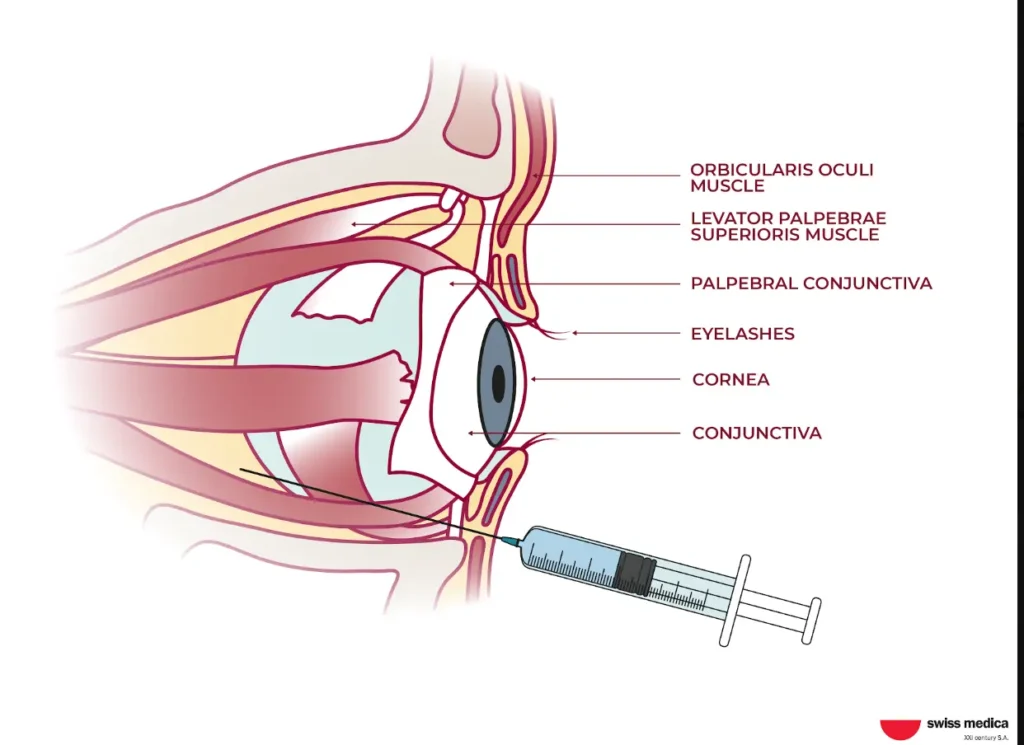
For retinal conditions, the cell-based part of the program may include intravenous infusion and retrobulbar administration, in which the product is delivered behind the eyeball. The exact route and treatment plan depend on the diagnosis, protocol, and physician’s evaluation.
Depending on the patient’s condition and the physician’s assessment, the program may combine cell-based treatment with additional therapies, including physiotherapy, Intracellular Metabolism Recovery (IMR), and exosome nasal drops.
They are selected individually after assessment, and the exact composition of the program is confirmed for each patient.
Follow-Up
Patients who undergo stem cell therapy for retinitis or retinopathy are monitored after treatment and remain in contact with the medical team through remote follow-up so that any changes can be assessed over time.
A free follow-up visit with accommodation is typically offered within 3–6 months. At this stage, doctors can review the patient’s progress and compare new findings with the pre-treatment baseline.
What Should Be Tracked After Treatment?
Changes after treatment should be evaluated using both objective eye tests and the patient’s own experience of any difference in vision.
OCT and Fundus Imaging Help Track Structural Changes
OCT and fundus photography provide a record of how retinal structures change over time. Doctors can use them to assess retinal thickness and monitor the retinal pigment epithelium (RPE). These findings provide objective information that may not always correspond directly to what the patient notices in daily life.
Visual Field Testing, ERG, and Contrast Sensitivity Can Detect Functional Changes
Visual field testing, electroretinography (ERG), and contrast sensitivity testing can reveal changes in retinal function that patients may not be able to describe precisely. Results can be compared with the patient’s pre-treatment baseline to track changes over time.
Safety & Side Effects and Evidence Limitations
Safety in stem cell treatment for retinitis and other retinal conditions depends not only on the cell product itself but also on patient selection, laboratory quality control, and the route of administration.
Possible Side Effects of Stem Cell Therapy
Possible short-term reactions after MSC-based therapy may include:
- a mild temporary rise in temperature;
- transient fatigue;
- local discomfort after administration.
These reactions are generally short-lived, and patients are monitored throughout recovery.
Read about stem cell therapy safety and side effects, published adverse-event data, and the laboratory and procedural controls used to reduce treatment risks at Swiss Medica.
Cost of Stem Cell Therapy for Retinitis and Retinopathy
At Swiss Medica, the cost of stem cell therapy for retinitis pigmentosa or diabetic retinopathy typically falls within a €7,700–€45,000* range. The exact stem cell therapy cost can be provided once doctors have assessed the patient’s condition and identified an appropriate treatment program.
Why Treatment Costs Depend on the Diagnosis and Treatment Plan
Factors that may influence the final cost include:
- diagnosis and disease stage;
- remaining visual function;
- retinal imaging and other tests required;
- cell type, source, and dosage;
- route of administration;
- number of treatment sessions;
- monitoring requirements;
- supportive therapies;
- accommodation and length of stay;
- follow-up.
These factors are reviewed before the treatment program, and final estimates are confirmed.
What a Swiss Medica Retinal Treatment Program May Include
The price alone does not show the full scope of a retinal treatment program. Depending on the provider, the quoted amount may refer to one procedure or a broader package that combines treatment with medical supervision and other services.
| Country / region | Approximate advertised price | What may be included |
| Mexico | $14,300–$30,200 per program | A single-clinic retinal protocol; accommodation and extended follow-up vary by provider |
| Turkey | $5,000–$10,000 per eye | Standard vs. premium retinal-dystrophy protocols; package contents differ by hospital tier |
| Germany | €20,800–€28,000 | Specialized regenerative/MSC-based eye programs; diagnostics and hospital fees may be priced separately |
| United States | $9,000–$16,000 per procedure | Retinitis pigmentosa-specific estimates, usually self-pay; follow-up beyond the immediate post-op period often not included |
| Serbia (Swiss Medica) | €7,000–€45,000* | A private full-hospital program with doctor monitoring, on-site accommodation, tailored meals, and client support |
At Swiss Medica, the final price is based on the treatment plan approved for the individual patient rather than on a fixed price per injection. The program combines cell-based treatment with medical monitoring, supportive care, accommodation, meals, and follow-up, depending on what is included in the patient’s plan. Travel to Serbia is arranged separately by the patient.
*Prices are indicative and based on 2026 estimates; they may vary depending on condition severity and required cell quantity.
Why Patients Choose Swiss Medica for Retinal Disease Treatment
Patients considering stem cell treatment for retinitis often come with a long history of ophthalmic examinations, ongoing treatment, and changes in vision over time. The same is true for many patients with other retinal diseases.
Individual Ophthalmic Assessment Before Treatment
Patients do not need to travel to Swiss Medica clinic simply to find out whether their case can be considered. Before the visit, doctors review available ophthalmology reports, previous treatment, and eye tests such as OCT, fundus imaging, ERG, and visual field results.
This review helps the medical team:
- understand the current condition of the retina and how the disease has progressed;
- see how much visual function remains;
- take previous retinal treatment and its results into account;
- decide whether further evaluation or regenerative treatment may be considered;
- discuss which treatment goals may be realistic and which expectations would not be.
This gives patients a clearer medical picture before making travel arrangements.
The same process applies to other eye and optic nerve conditions. Read about stem cell therapy for glaucoma to see how candidacy is assessed there.
In-House Cell Processing and Quality Standards
For patients, laboratory control means knowing that the cell product has been checked before it is used. Swiss Medica’s laboratory works to EU GMP Grade A cleanliness standards and is regularly audited.
At Swiss Medica, cell products are prepared and tested in-house rather than being supplied by an external laboratory and transported between separate facilities. Keeping preparation and treatment within the same center helps avoid unnecessary transport and handling and allows the medical team to follow the product from preparation to administration.
Before treatment, each batch is checked for sterility, identity, and viability. This helps confirm that the product is free from contamination, contains the expected cell type, and has a sufficient proportion of living cells.
These controls do not guarantee a treatment result, but they give patients a more transparent and controlled quality process before the cells are administered.
Personalized Monitoring
For retinal disease, follow-up is most informative when new findings are compared with the patient’s own pre-treatment baseline.
Depending on the diagnosis and the tests available, doctors may review changes in OCT or fundus images together with visual field, ERG, contrast sensitivity, and the patient’s own experience. This can help put reported changes into context and give the patient and their local ophthalmologist clearer information for continued retinal care.
International Patient Support in Serbia
Traveling abroad can be more demanding when vision is limited. Navigating an unfamiliar airport or clinic, arranging transport, and managing everyday practical details can add extra stress for both patients and the people traveling with them.
At Swiss Medica’s Belgrade hospital, the care setting is organized to make the stay easier for international patients:
- treatment and accommodation are provided in one location, reducing the need to travel between facilities;
- medical staff are available 24/7 throughout the stay;
- practical and logistical support helps patients spend less time organizing everyday arrangements;
- the hospital provides a comfortable setting for rest and recovery between treatments;
- facilities are adapted for patients with limited mobility and other accessibility needs.
This allows patients to focus more fully on their treatment and recovery while the practical aspects of their stay are supported.
Learn more about Swiss Medica, including our history, modern hospitals, in-house laboratory, and approach to regenerative medicine.



Speak With a Medical Advisor About Retinal Disease Treatment Options
If retinitis pigmentosa, diabetic retinopathy, or another retinal condition continues to affect your vision despite standard care, it can be difficult to know whether regenerative treatment is worth considering. A free confidential online consultation can help you better understand your options and whether stem cell therapy may be appropriate to discuss in your case.
Contact us
Send us your recent eye records, and the medical team will review your diagnosis and test findings, explain what outcomes may realistically be expected, and advise whether further evaluation or a treatment program may be appropriate.
Medical Advisor, Swiss Medica doctor
FAQ
1. Can stem cells repair retinal damage?
Current evidence does not show that stem cells can repair established retinal damage. MSCs are being studied for their potential to regulate inflammation and support the retinal environment and surviving tissue, but reproducible regeneration of lost photoreceptors has not been demonstrated. This is therefore being studied as a supportive approach rather than a way to rebuild lost retinal tissue.
2. Can stem cell therapy help retinitis pigmentosa?
Some human studies have reported stabilization of visual field measures and retinal structure, while results for visual acuity have been mixed. The evidence is still limited, and larger controlled studies are needed to determine which patients may benefit and how durable any improvements are.
3. Can stem cells help diabetic retinopathy?
For diabetic retinopathy, stem cell therapy remains largely at the preclinical research stage. Blood sugar control and standard treatments such as anti-VEGF injections or laser therapy remain the foundation of care. At present, stem cell therapy should be viewed as an additional option rather than a replacement for established treatment.
4. Can stem cell therapy restore lost vision?
Current evidence does not show that stem cell treatment for retinitis or retinopathy can restore vision that has already been lost. Published studies have more often reported stabilization of remaining visual function or modest improvements in specific measures, but these results should not be interpreted as reversal of established vision loss.
5. Is retinal stem cell therapy proven?
Cell-based therapy for retinal diseases is still investigational and has not been established as standard medical care. Studies in retinitis pigmentosa have reported encouraging safety results along with some functional improvements. Larger controlled trials are still needed, however, to determine how effective the treatment may be and which patients are most likely to benefit.
6. Who may be reviewed for stem cell treatment for retinitis or retinopathy?
Patients with retinitis pigmentosa, diabetic retinopathy, or other inherited or degenerative retinal disorders may be considered for individual review, particularly when the condition continues to progress despite standard care. Suitability depends on the diagnosis, disease stage, remaining retinal function, imaging findings, medical history, and overall safety profile. The decision is made after medical record review and individual assessment.
7. Does stem cell therapy replace anti-VEGF injections, laser, or surgery?
No. Stem cell therapy is being studied as an addition to standard retinal care, not as a substitute for it. Anti-VEGF injections, laser treatment, surgery when indicated, and diabetes management remain established approaches for their respective conditions. Patients should not stop or delay recommended standard treatment on the assumption that stem cell therapy can replace it.
8. What are the risks of stem cell therapy for retinal disease?
Temporary discomfort at the treatment site, short-lived inflammation, mild fever, or fatigue may occur after treatment. The reactions a patient may experience depend on the cell product, administration route, and procedure used. Proper laboratory standards, medical screening, and post-treatment monitoring therefore remain important.
9. How much does retinal stem cell therapy cost?
At Swiss Medica, treatment programs typically range from €7,700 to €45,000*. The exact price is determined after medical review and depends on factors such as the diagnosis and stage of the condition, required imaging, cell type and dosage, administration route, number of sessions, supportive care, accommodation, and follow-up. Each patient receives an individual cost estimate based on their records and the treatment plan recommended for their case.
* Prices are indicative 2026 estimates and vary by case; a personalized figure follows medical record review.
10. What eye tests are needed before consultation?
For the initial review, recent OCT scans, fundus photographs, ERG results, and visual field tests are especially useful. Patients can also provide fluorescein angiography and previous ophthalmology reports if available. Earlier records may be helpful as doctors need to see how the retinal condition has progressed over time.
References:
O’Neal TB, Tripathy K, Luther EE. Retinitis Pigmentosa. In: StatPearls [Internet]. Treasure Island (FL): StatPearls Publishing; updated 2024 Feb 12. PMID: 30137803.
Shukla UV, Tripathy K. Diabetic Retinopathy. In: StatPearls [Internet]. Treasure Island (FL): StatPearls Publishing; updated 2023 Aug 25. PMID: 32809640.
Drag S, Dotiwala F, Upadhyay AK. Gene Therapy for Retinal Degenerative Diseases: Progress, Challenges, and Future Directions. Invest Ophthalmol Vis Sci. 2023;64(7):39. doi:10.1167/iovs.64.7.39. PMID: 37389545. PMCID: PMC10318594.
Kąpa M, Koryciarz I, Kustosik N, Jurowski P, Pniakowska Z. Future Directions in Diabetic Retinopathy Treatment: Stem Cell Therapy, Nanotechnology, and PPARα Modulation. J Clin Med. 2025;14(3):683. doi:10.3390/jcm14030683. PMID: 39941353. PMCID: PMC11818668.
Krungkraipetch L, Supajitgulchai D, Assawaboonyadech A, Puranawit W. Efficacy and safety of mesenchymal stem cell therapies in retinitis pigmentosa: a systematic review and meta-analysis. Int Ophthalmol. 2025;45(1):85. doi:10.1007/s10792-025-03478-6. PMID: 40072800.
Tuekprakhon A, Sangkitporn S, Trinavarat A, et al. Intravitreal autologous mesenchymal stem cell transplantation: a non-randomized phase I clinical trial in patients with retinitis pigmentosa. Stem Cell Res Ther. 2021;12(1):52. doi:10.1186/s13287-020-02122-7. PMID: 33422139. PMCID: PMC7796606.
Soltani Khaboushan A, Ebadpour N, Johari Moghadam MM, Rezaee Z, Kajbafzadeh AM, Majidi Zolbin M. Cell therapy for retinal degenerative disorders: a systematic review and three-level meta-analysis. J Transl Med. 2024;22(1):227. doi:10.1186/s12967-024-05016-x. PMID: 38431596. PMCID: PMC10908175.
Gu X, Yu X, Zhao C, et al. Efficacy and Safety of Autologous Bone Marrow Mesenchymal Stem Cell Transplantation in Patients with Diabetic Retinopathy. Cell Physiol Biochem. 2018;49(1):40–52. doi:10.1159/000492838. PMID: 30134223.
Sarsenova M, Alimbek S, Imanbekova M, et al. Diabetic Retinopathy and Current Treatment Approaches: From Pathogenesis to Extracellular Vesicle-Based Therapies. Int J Mol Sci. 2026;27(14):6354. doi:10.3390/ijms27146354. PMID: 42511696. PMCID: PMC13411447.
Wang Y, Yi H, Song Y. The safety of MSC therapy over the past 15 years: a meta-analysis. Stem Cell Res Ther. 2021;12(1):545. doi:10.1186/s13287-021-02609-x. PMID: 34663461. PMCID: PMC8522073.
Qi XY, Mi CH, Cao DR, Chen XQ, Zhang P. Retinitis pigmentosa and stem cell therapy. Int J Ophthalmol. 2024;17(7):1363–1369. doi:10.18240/ijo.2024.07.22. PMID: 39026924. PMCID: PMC11246939.


MD, Pediatrician, Regenerative Medicine Specialist